Spectrometer:
The SPECs electron spectrometer used is a standard Phoibos 150 elevated pressure. The spectrometer is fitted with a PCO.edge 5.5 and a 35mm focal length Navitar lens. The camera is operated at the FEL repetion rate, capturing an image of the detector for each individual FEL events. The images are tagged to the proper pulse ids allowing the correlation with other detectors/spectrometers in the machine and endstation.
The spectrometer is typically operated in Fixed Analyzer Transmission (FAT) mode. In this mode, the spectrometer does not sweep its energy, but resolved a fixed energy window of the electrons with a fixed central kinetic (Ek) and pass energy (Ep). The Ek energy defines the central energy of the resolved spectra, while Ep defines the width of the width of the window. A compromise between the width of the energy window, resolution, and transmission is balanced based on requirements.
The spectrometer could be operated in a variety of operation modes. Each mode offers different energy resolution, transmission, spatial imaging, and angle resolution.
Example 1: For single shot spectral characterization we use Medium Area (MA) mode as it images an area ~2x7mm in the interaction point and offers high electron transmission. Further this mode can reach up to Ek=1KeV and Ep=280eV offering a window of 35eV.
Example 2: For spectroscopy, Medium Angle Mode (MAM) mode offers high Ep energies up to 500eV allowing large energy windows up to 100eV. This mode images a smaller area than the MA mode, offering better spatial sampling of the interaction point.
Image Correction: Image transformation, correction, and calibration is required to produce spectra. An image transform script is prepared that performs the corrections and produce the spectra on shot-to-shot basis.
Example: MAM mode, Ek=72eV, Ep=320eV probing CO2 electrons with 540eV x-rays.
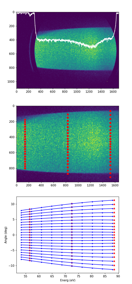
- First image: Integration over 1000 FEL events (required as we record 50e-/shot)
- Second image: Image crop and resize. Red dots are anchor points for the transformation to warp.
- Third image: Interpolation between the anchor points, allowing smooth transformation process.
A configuration setting is required for each operation mode, Ek and Ep that is offered by the SPECs software. The configuration file would look as follows:

This script offers a calibrated and transmission corrected electron spectra for each individual image inserted.